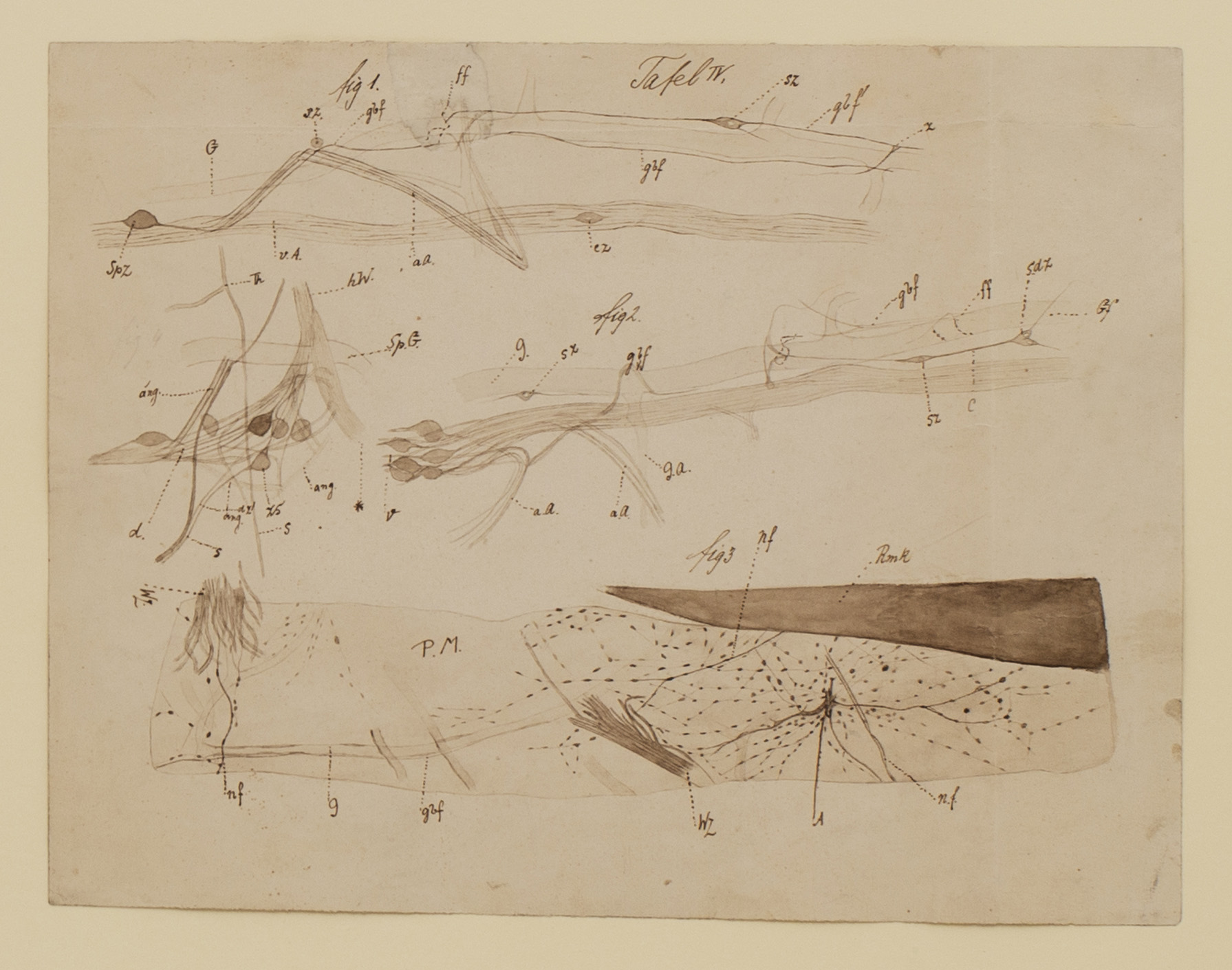
The drawing is an aggregate of various cross-sections and staining of the Petromyzon.
In Figure 1, we see a posterior view of the Petromyzon’s nerves.
The spinal ganglionic cell (spz), an archaic aggregation of nerve cells, is located in the left corner.
Freud includes many elements of the nervous system, while following one nerve fiber as it accompanies a microscopic blood vessel (gbf).
Freud also illustrates a varicose axon of a nerve cell (ff), a part of a nerve fiber not coated with any other cells, and thus with a lower conductivity level.
The capacity to see nerve cells with such a high level of precision was one of the most novel elements in nerve histology and was the result of a staining technique developed by the Spanish neurobiologist Cajal.
Figure 2 depicts a different branch of the posterior root of the spinal ganglion of the Petromyzon.
Of particular note are the black elements, marked as zs, representing spinal ganglion cells firing into the sympathetic branch of the Petromyzon’s nerve system.
On the bottom of the drawing, Figure 3 shows a network of nerve cells attached to the pia mater, one of the nerve’s meninges, or coating, marked in drawing as PM.
On both the right and left hand side, branches of nerve fibers (nf) spread out of the spinal cord (marked Rmk) through a larger plexus of nerve fibers (A).